Open source tools for vascular physiology

Open source pressure myograph

VasoTracker is a collection of open source tools for studying vascular physiology.  We started off with a simple bath for pressure myography, but we are constantly working on new things. So far, our collection includes: a pressure myograph, diameter tracking software, temperature controller, and a pressure monitor.

Open Source Pressure Myography

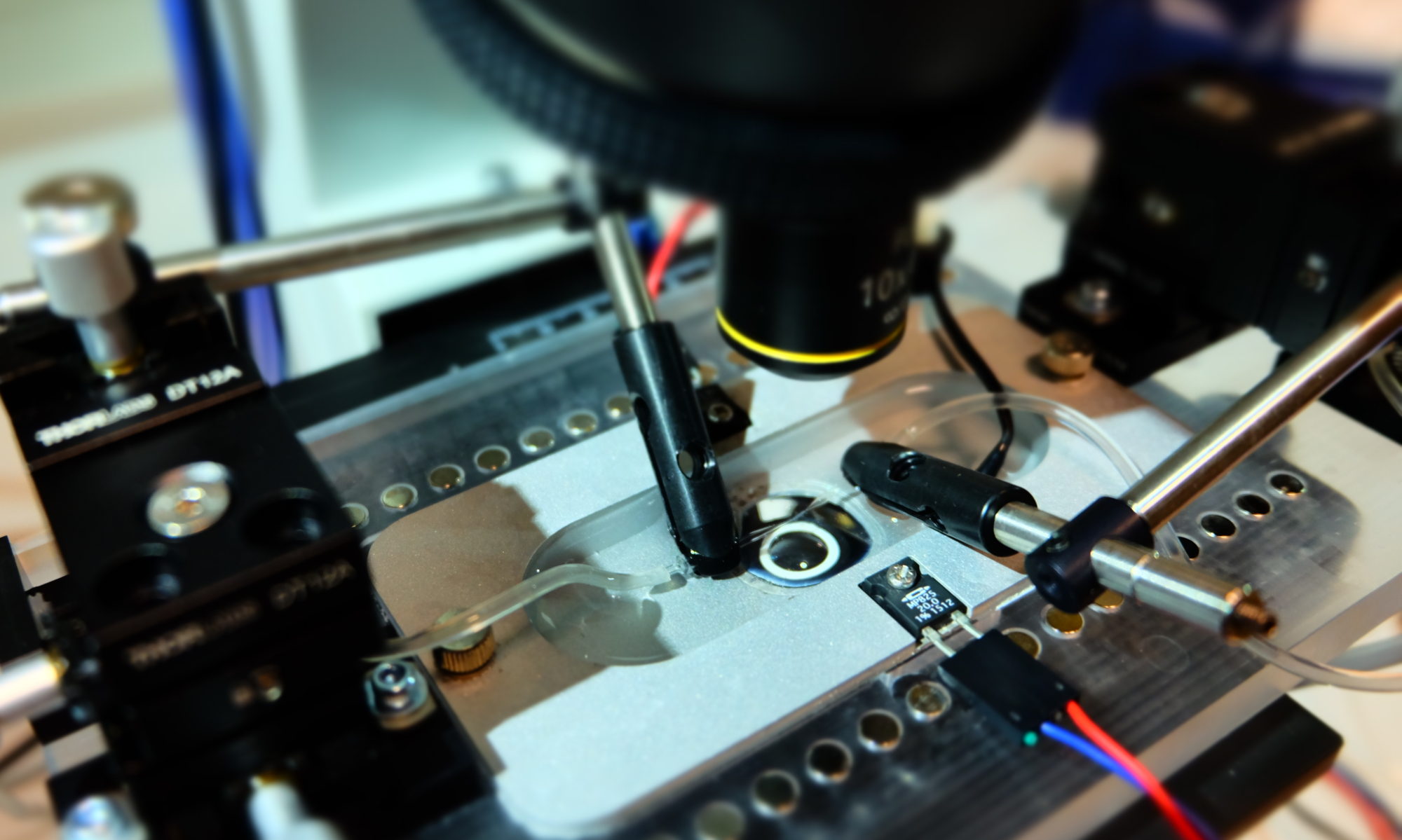
Pressure myography is the gold standard for measuring blood vessel function. These systems allow us to study the effects of drugs and other compounds on arteries, veins, and other vessels. The technique also allow an evaluation of blood vessel responses to mechanical  stimuli, such as increases in pressure or flow. So whether your lab is interested in assessing smooth muscle cell contraction or endothelium-dependent vasodilation, the pressure myograph is an essential piece of equipment.

A while ago, we wanted to set up a new pressure myograph (check us out here). We knew that there are a number of different commercial myograph packages available. However, the cost of even the most primitive systems took us by surprise. A little frustrated, and unwilling to part with so much money, we decided to develop our own, VasoTracker.

The VasoTracker pressure myograph is robust and it works very well. Best of all, the complete system cost us less than 10% of the equivalent from a leading commercial suppliers (we won’t say who!). We thought that others might also like to benefit from VasoTracker, so we decided to make everything (our hardware designs, our software, and instructions for setting everything up) freely available. It is also open source,  which means that we have documented the system in detail and users are free to modify it in any way.

We announced the birth of the VasoTracker pressure myograph in Frontiers in Physiology. More details on our system can be found on our here.

Live Diameter Tracking Software

In pressure myography, the main physiological readout of artery function is diameter. The system uses video microscopy techniques to track the diameter of pressurized blood vessels. We wrote our own diameter tracking software. And we are very proud of it. You can find more details here.

Offline Diameter Tracking Software

Sometimes we just record data, and worry about analyzing it later. If this sounds like you, you might want to take a look at VasoTrackerOA (OA stands for offline analyzer). VasoTrackerOA is a piece of software that loads in video files (tiff stacks, avi or wmv files) and measures diameter. We have tested it on a variety of data from pressure myography, vascular ultrasound, and fluorescence imaging experiments. Details are here.

The video below shows our VasoTracker software in action. Blue lines show the outer diameter of the artery. Red lines show the inner diameter.  Check out our software page for more details.

Citing VasoTracker

Support our work

VasoTracker is a collection of open source hardware and software – it was made possible by grant funding from the Wellcome Trust and British Heart Foundation. Our ability to support, develop and maintain VasoTracker will depend on more grants, so whether you use a complete VasoTracker system, just some of the components, or the software, please cite our work in your scientific publications.

  • Lawton, P. F., Lee, M. D., Saunter, C. D., Girkin, J. M., McCarron, J. G., & Wilson, C. (2019). VasoTracker, a Low-Cost and Open Source Pressure Myograph System for Vascular Physiology. Frontiers in Physiology, 10. DOI: 10.3389/fphys.2019.00099

Papers citing VasoTracker

  1. Rodríguez-Rodríguez, R., Ackermann, T. N., Plaza, J. A., Simonsen, U., Matchkov, V., Llobera, A., & Munoz-Berbel, X. (2019). Ultrasensitive photonic microsystem enabling sub-micrometric monitoring of arterial oscillations for advanced cardiovascular studies. Frontiers in Physiology. DOI: 10.3389/fphys.2019.00940
  2. Heathcote, H. R., Lee, M. D., Zhang, X., Saunter, C. D., Wilson, C., & McCarron, J. G. (2019). Endothelial TRPV4 channels modulate vascular tone by Ca2+‐induced Ca2+ release at inositol 1,4,5‐trisphosphate receptors. British Journal of Pharmacology. DOI: 10.1111/bph.14762
  3. Wilson, C., Zhang, X.,Buckley, C., Heathcote, H. R., Lee, M. D., & McCarron, J. G. (2019). Increased vascular contractility in hypertension results from impaired endothelial calcium signaling. Hypertension. DOI: 10.1161/HYPERTENSIONAHA.119.13791
  4. Edwards, J. M., Roy, S., Tomcho, J. C., Schreckenberger, Z. J., Chakraborty, S., Bearss, N. R., … Wenceslau, C. F. (2019). Microbiota are critical for vascular physiology: Germ-free status weakens contractility and induces sex-specific vascular remodeling in mice. Vascular Pharmacology. DOI: 10.1016/j.vph.2019.106633 [Okay, well they didn’t cite us, but they did use VasoTracker]
  5. Barabas, P., Augustine, J., Fernández, J. A., McGeown, J. G., McGahon, M. K., & Curtis, T. M. (2020). Ion channels and myogenic activity in retinal arterioles. In Current Topics in Membranes (pp. 187–226). Elsevier. DOI: 10.1016/bs.ctm.2020.01.008
  6. Castorena‐Gonzalez, J. A., Srinivasan, R. S., King, P. D., Simon, A. M., & Davis, M. J. (2020). Simplified method to quantify valve back‐leak uncovers severe mesenteric lymphatic valve dysfunction in mice deficient in connexins 43 and 37. The Journal of Physiology. DOI: 10.1113/jp279472
  7. Wilson, C., Zhang, X.,Buckley, C., Heathcote, H. R., Lee, M. D., & McCarron, J. G. (2019). Disrupted endothelial cell heterogeneity and network organization impair vascular function in prediabetic obesity. Metabolism. DOI: 10.1016/j.metabol.2020.154340
%d bloggers like this: